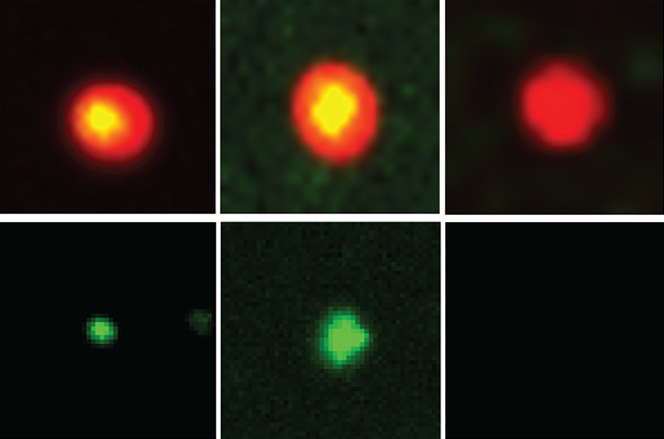
 Influenza A virus particles contain free, “unanchored” ubiquitin. Viral particles are shown in red, unanchored ubiquitin in green, and their overlap in yellow. Credits: Shi et al., Science Advances
September 28, 2026
New tool tracks a hidden protein involved in viral infection and cell cleanup
Ubiquitin is a small protein that usually works by attaching itself to other proteins. But it can also exist freely, on its own or in chains, and this form has been hard to study. FMI researchers and their collaborators have now developed a tool called UBiP10 that can pick out and reveal this free ubiquitin in cells and influenza A virus particles. Using it, they found that free ubiquitin gathers around protein aggregates, offering a new way to study its role in viral infection and the cell’s protein-cleanup systems.
Viruses such as influenza A and dengue can hijack a cellular system that helps manage protein aggregates. Previous work by FMI emeritus group leader Patrick Matthias and his team showed that a small protein called ubiquitin plays an important role in this process.
The viruses rely on a free form of ubiquitin that is not attached to other proteins. This “unanchored” ubiquitin is also involved in immune responses and protein cleanup, but it has been difficult to study because researchers lack tools that can specifically detect it.
So, Matthias and collaborators at ETH Zurich set out to build a tool that could pick out unanchored ubiquitin from other forms of the protein. Working with researchers in Guillaume Diss’s lab and the FMI structural biology facility, Longlong Wang — a former postdoc with Matthias — started with HDAC6, a protein that naturally binds unanchored ubiquitin. They improved the binding, then used computer-based protein design tools, including AlphaFold, to generate thousands of new versions.
One candidate, called UBiP10, performed especially well. When tested in the lab, it captured unanchored ubiquitin while mostly ignoring ubiquitin attached to other proteins. It could also isolate unanchored ubiquitin from human cells and influenza A virus particles.
The researchers then used UBiP10 to see where unanchored ubiquitin collects inside cells. When the proteasome — the cell’s main protein-disposal system — was blocked, damaged proteins gathered into structures called aggresomes. UBiP10 showed that unanchored ubiquitin formed a layer around these structures rather than building up inside them. This layer depended on a motor protein called myosin 10.
The role of this ubiquitin-rich layer is still unclear. But UBiP10 gives researchers a new way to track a form of ubiquitin that has been difficult to study, which could help clarify its roles in viral infection and the cell’s protein-cleanup systems, Matthias says. The team has also filed a patent application for the synthetic protein.
Original publication
Shihua Shi, Alexandra Bendel, Georg Kempf, Jacint Sanchez, Sohei Majima, Vytautas Iesmantavicius, Daniel Hess, Jan Seebacher, Yong Huang, Sucheta Ghosh, Chun Cao, Gabriele Matthias, Jeffrey Bode, Yohei Yamauchi, Guillaume Diss, Patrick Matthias* & Longlong Wang* A designed ubiquitin-binding protein selectively enriches and visualizes unanchored ubiquitin Science Advances (2026)
* co-corresponding authors
Influenza A virus particles contain free, “unanchored” ubiquitin. Viral particles are shown in red, unanchored ubiquitin in green, and their overlap in yellow. Credits: Shi et al., Science Advances


